
Rutgers CryoEM & Nanoimaging Facility
The Rutgers CryoEM & Nanoimaging Facility (RCNF) preserves biological specimens in their native state by vitrification; images them at the nanoscale through cryo-transmission electron microscopy, cryo-scanning electron microscopy, or cryo-focused ion beam microscopy; and reconstructs three-dimensional information through computational analysis of these nanoscale images. We changed our name from the Rutgers New Jersey Cryo-Electron Microscopy and Tomography Core Facility in October 2020 to reflect the diversity of imaging techniques employed in the facility.
The facility is available on a fee-for-use basis, with preference in scheduling and pricing to members of the Rutgers community. Potential users are welcomed to contact the director to discuss how cryoEM can be applied to their projects. Before bringing a specimen to the facility for cryoTEM, users should provide supporting data that their specimen is of a suitable concentration (generally, 0.2-1 mg/mL), suitable buffer composition, and of sufficient purity for cryo-electron microscopy.
Equipment
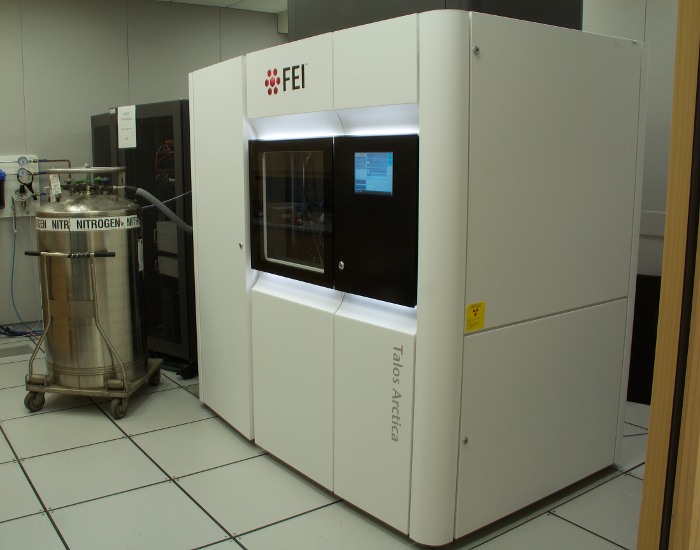
For single-particle reconstruction of protein complexes and for cryoelectron tomography, we image around the clock on our 200 kilovolt Talos Arctica cryogenic transmission electron microscope. It is equipped with a BioQuantum Gatan Image Filter to remove inelastically scattered electrons, a Volta phase plate for contrast enhancement, and a Summit K2 direct electron detector for optimal detector performance at high resolution. This configuration is equally suitable for single-particle and tomographic studies. The instrument is piloted remotely from an adjoining control room (or over the ‘net) and is outfitted with automated collection software.
Vitrification by plunge-freezing is achieved with a Leica EM-GP plunge freezer, capable of front- or back-blotting, or with a Vitrobot Mark IV dual-blotting plunger. Plunge freezers generate an aqueous layer of reproducible thickness across the EM grid and rapidly chill this layer to an amorphous, solid state. The EM-GP is ideal for cell studies and also creates uniform-thickness ice for single-particle work. We stock standard grid styles and have a Pelco EasiGlow available for hydrophilization.
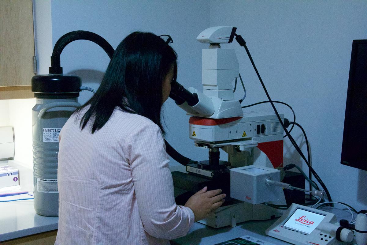
Correlative light microscopy/electron microscopy (CLEM) experiments are conducted on a Leica DM6 FS/EM Cryo CLEM system. After vitrifying a sample on an electron microscopy grid, the same grid can be imaged in fluorescence (or brightfield) optical microscopy and in the cryoelectron microscope. Fiducial markers seen in both optical and electron microscopy are used to correlate the images. In one embodiment, a quick scan of the grid by optical microscopy is used to locate a rare event, and the user tracks the electron microscope stage to the correct site to record a tomogram or mill a lamella at that location.
In 2020, we installed North America’s first Aquilos 2 cryogenic focused ion beam/scanning electron microscope. The cryoFIB/SEM is used to micromachine lamellae from cells to prepare specimens for in situ cryoelectron tomography, to perform serial section imaging by ablating a few nanometers of material at a time between SEM images, or to mill bits of tissue for lift-out cryoTEM preparation. The instrument can be used at room temperature as well, but to maintain an ideal state for all users, we require that all specimens go in and out through the airlock regardless of temperature.
For surface imaging, our LEO 1525 field emission gun scanning electron microscope is equipped with at Gatan Alto 2500 cryostage. The Alto adds slush-freezing, controlled sublimation/freeze-etch, and on-column sputter coating to the FEG-SEM. This instrument is very easy to use despite its high resolution. For room-temperature preparation, a Leica ACE600 electron beam deposition unit can spray ultrathin metallic platinum or carbon in coating or low-angle rotary shadowing experiments.
We’ve partnered with the Office of Advanced Research Computing to maintain a university-wide installation of certain cryoEM software on the Amarel cluster. Contact the director for further details. The facility maintains some resources for data processing in-house on a buy-in basis.
Useful Links
Cryoelectron microscopy is complementary to classical electron imaging techniques such classical transmission electron microscopy. The Core Imaging Lab, a unit of the Robert Wood Johnson Medical School Department of Pathology, provides services such as TEM of stained or sectioned samples to the Rutgers community.
The research interests of several members of the Institute for Quantitative Biomedicine include structural biology generally or cryo-electron microscopy techniques. In particular, Professors Wei Dai, Arek Kulczyk , and Vasileios Petrou are leaders in cryo-electron tomography and cryo-electron microscopy at Rutgers. For example, the Kulczyk lab is developing new techniques in single-molecule CLEM as well as applying high-resolution single-particle analysis to biological targets. The Petrou lab has especial expertise in single-particle cryoEM of membrane proteins. The Dai lab integrates cryo-electron tomography with CLEM and other tools to study nanomachines and disease-associated structures in their native cellular context, and has pioneered application of phase plates in biological electron microscopy.
Contact information
The facility is located on the ground floor of the Proteomics building, which we share with the high-field NMR and biological mass spectrometry facilities. The facility director, Prof. Jason Kaelber, can be contacted by email.